
Diabetic retinopathy is a leading cause of vision loss among working-age adults in the United States, affecting millions of Americans, with the CDC estimating on the order of 8 million people living with some degree of the disease. Diabetic retinopathy occurs when chronically elevated blood sugar damages the tiny blood vessels in the retina, the light-sensitive tissue at the back of the eye. What makes this condition particularly insidious is that it often produces no symptoms in its early stages, silently progressing until significant, sometimes irreversible, vision loss has occurred. Regular eye exams and tight blood sugar control are your best defenses. This guide covers the full spectrum from early detection to advanced treatment. For other diabetes complications, see our medical conditions guide.
TL;DR: What to Know
- Silent early on: early diabetic retinopathy usually has no symptoms, so regular dilated eye exams, not how your vision feels, are what catch it in time.
- Stages: nonproliferative (NPDR) is earlier; proliferative (PDR), with fragile new blood vessels, is advanced and vision-threatening. Diabetic macular edema can occur at any stage.
- Control the basics: blood sugar, blood pressure, cholesterol, and not smoking protect your retinas at every stage.
- Modern treatment works: anti-VEGF injections, laser, and surgery can preserve and sometimes improve vision; newer longer-acting injections can reduce treatment frequency.
- Know the emergencies: sudden vision loss, many new floaters, flashes, or a curtain or shadow across your sight need urgent eye care (see the red-flag box).
This article is general education, not medical advice. Diagnosis and all treatment should be directed by an ophthalmologist or your diabetes care team.
How Diabetes Affects the Eyes
The retina is one of the most metabolically active tissues in the body and depends on a dense network of small blood vessels for its oxygen supply. Chronic hyperglycemia damages the endothelial cells lining these vessels through several mechanisms: oxidative stress, accumulation of advanced glycation end products, and activation of inflammatory pathways. The result is a progressive weakening of the retinal vasculature.
As retinal blood vessels are damaged, they may leak fluid and blood into the retina, become blocked (leading to retinal ischemia), or grow abnormally in a desperate attempt to restore blood supply. These changes, leakage, ischemia, and neovascularization, underlie the different stages and complications of diabetic retinopathy. The duration of diabetes is the strongest risk factor; after 20 years of diabetes, a large majority of people with type 1 and many with type 2 diabetes will have some degree of retinopathy, according to the National Eye Institute.
Diabetic retinopathy does not exist in isolation. It frequently coexists with other diabetes complications, including diabetic neuropathy and diabetic kidney disease, reflecting the systemic nature of diabetic microvascular damage. The same risk factors, poor glycemic control, hypertension, high cholesterol, and smoking, accelerate all of these conditions.
Stages of Diabetic Retinopathy
Nonproliferative Diabetic Retinopathy (NPDR)
NPDR is the earlier stage and is classified as mild, moderate, or severe based on the findings on a dilated eye exam. In mild NPDR, small areas of balloon-like swelling (microaneurysms) form in the retinal blood vessels. These may leak small amounts of fluid but typically do not affect vision. As NPDR progresses to moderate and severe stages, more vessels become damaged and blocked. Cotton-wool spots (areas of retinal ischemia), venous beading, and intraretinal microvascular abnormalities (IRMA) appear. Severe NPDR indicates widespread retinal ischemia and carries a high risk of progressing to the proliferative stage.
Proliferative Diabetic Retinopathy (PDR)
PDR is the advanced stage in which ischemia triggers the growth of new, abnormal blood vessels (neovascularization) on the surface of the retina or optic disc. These new vessels are fragile and prone to bleeding into the vitreous cavity (vitreous hemorrhage), which can cause sudden, severe vision loss. Neovascular tissue can also form scar tissue that contracts and pulls on the retina, leading to tractional retinal detachment, one of the most serious complications of diabetic retinopathy. PDR is a vision-threatening condition that requires prompt treatment by a retina specialist.
Diabetic Macular Edema (DME)
Diabetic macular edema can occur at any stage of retinopathy and is the most common cause of vision loss in people with diabetic retinopathy. It occurs when damaged blood vessels leak fluid into the macula, the central part of the retina responsible for sharp, detailed vision. The swelling distorts vision, causing blurriness, difficulty reading, and impaired color perception. DME affects hundreds of thousands of Americans and is a major focus of modern treatment, according to research summarized by the American Academy of Ophthalmology and published in journals such as Ophthalmology.
Symptoms and When They Appear
In the early stages, diabetic retinopathy usually causes no symptoms at all. This is the central challenge, by the time you notice vision changes, significant retinal damage may already have occurred. This reality underscores the critical importance of regular dilated eye exams for all people with diabetes, regardless of whether their vision seems fine.
As the disease progresses, symptoms may include blurred or fluctuating vision, dark spots or “floaters” (caused by blood leaking into the vitreous), difficulty seeing at night, faded or washed-out colors, and dark or empty areas in your field of vision. A sudden shower of floaters, flashes of light, or a curtain-like shadow over your vision may indicate a vitreous hemorrhage or retinal detachment and requires immediate medical attention.
Diabetic macular edema may cause central vision blurriness that makes reading, driving, and recognizing faces difficult. Some patients notice that straight lines appear wavy or distorted. These symptoms can develop gradually or relatively quickly, depending on the rate of fluid leakage.
Red Flags: Seek Urgent Eye Care
Some symptoms can signal a bleed or a detaching retina and need same-day evaluation. Contact an ophthalmologist immediately or go to the emergency room if you experience:
- Sudden loss of vision in one or both eyes, even if it is painless
- A sudden increase in floaters or a “shower” of new spots or specks
- Flashes of light that are new or persistent
- A shadow, curtain, or dark area spreading across your field of vision
These may indicate a vitreous hemorrhage or retinal detachment. Prompt treatment offers the best chance of saving vision, so do not wait for a routine appointment.
Risk Factors and Prevention
The duration of diabetes is the most powerful predictor of retinopathy. The longer you have had diabetes, the higher your risk. Poor glycemic control accelerates the timeline; the landmark DCCT trial demonstrated that intensive blood sugar management in type 1 diabetes substantially reduced the risk of developing retinopathy and slowed its progression. In type 2 diabetes, the UKPDS trial showed similar benefits of tight glucose control.
Hypertension is a major independent risk factor. High blood pressure damages retinal blood vessels and exacerbates the effects of hyperglycemia. The UKPDS also demonstrated that tight blood pressure control significantly reduced the risk of retinopathy progression. Current American Diabetes Association guidance generally recommends a blood pressure target below 130/80 mmHg for many people with diabetes, individualized to the patient.
Other risk factors include high cholesterol and triglycerides (which contribute to hard exudates, lipid deposits in the retina), pregnancy (which can accelerate retinopathy progression, so a dilated exam is recommended during pregnancy planning and each trimester), smoking, and kidney disease. Interestingly, a rapid improvement in blood sugar control can temporarily worsen retinopathy in some patients, a phenomenon called “early worsening,” which is why eye exams are recommended before and after significant changes in diabetes management, including when starting newer medications that lower blood sugar quickly.
Screening and Diagnosis
The American Diabetes Association, in its annually updated Standards of Care in Diabetes, recommends the following general screening schedule: for type 1 diabetes, an initial dilated eye exam within five years of diagnosis, then at least annually; for type 2 diabetes, an initial exam at the time of diagnosis, then at least annually. If there is no retinopathy on one or more annual exams and blood sugar is well controlled, screening intervals may be extended to every one to two years at the discretion of the eye care provider. Because these standards are refreshed each year, confirm the current recommendation with your care team.
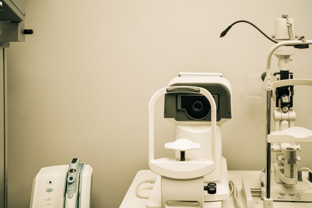
The comprehensive dilated eye exam is the standard screening method. Dilating drops widen the pupil, allowing the ophthalmologist or optometrist to examine the retina thoroughly. Retinal photography and optical coherence tomography (OCT), which provides cross-sectional images of the retina, are increasingly used as adjuncts. OCT is particularly valuable for detecting and monitoring macular edema, and OCT angiography can map retinal blood flow without dye.
Fluorescein angiography, in which a fluorescent dye is injected into the bloodstream and photographed as it flows through the retinal vessels, is used to identify areas of leakage, blockage, and neovascularization. This test is typically reserved for patients with known retinopathy who are being evaluated for treatment. Autonomous, artificial-intelligence-based screening systems that analyze retinal photographs are FDA-cleared and now deployed in primary care and pharmacy settings, allowing some patients to be screened for referable retinopathy during a routine visit without a specialist present. These tools improve access, especially in areas with limited eye care, though a positive or ungradable result still leads to a full eye exam.
Treatment for Nonproliferative Retinopathy
Mild to moderate NPDR without macular edema generally does not require eye-specific treatment. The focus is on optimizing systemic risk factors: achieving and maintaining an individualized A1C target (often around 7 percent for many adults), controlling blood pressure, managing lipids, and avoiding smoking. Regular monitoring, typically every 6 to 12 months, allows early detection of progression.
For diabetic macular edema, anti-vascular endothelial growth factor (anti-VEGF) injections are the current standard of care. These medications, which include ranibizumab (Lucentis), aflibercept (Eylea), bevacizumab (Avastin, used off-label), and newer agents such as faricimab (Vabysmo) and high-dose aflibercept 8 mg (Eylea HD), are injected directly into the vitreous cavity. They work by blocking the growth factor that drives abnormal blood vessel leakage and growth. Clinical trials published in the New England Journal of Medicine have shown that anti-VEGF therapy not only stabilizes vision but often improves it, with some patients gaining two or more lines on the eye chart.
Anti-VEGF injections are typically given monthly at first, with the interval gradually extended based on response. A significant recent development is the arrival of longer-acting options: faricimab and high-dose aflibercept are designed to maintain results with less frequent injections for many patients, and biosimilar versions of older anti-VEGF drugs have expanded lower-cost choices. Most patients still require ongoing treatment for years, so adherence to follow-up is essential. Intravitreal corticosteroid implants (such as dexamethasone or fluocinolone acetonide) are alternatives for patients who do not respond adequately to anti-VEGF therapy or who prefer less frequent dosing, though they carry risks of cataract and glaucoma. The specific drug, dose, and schedule are always chosen and administered by your retina specialist.
Treatment for Proliferative Retinopathy
Proliferative diabetic retinopathy requires more aggressive intervention. Panretinal photocoagulation (PRP), also known as scatter laser treatment, has been a mainstay of PDR treatment for decades. PRP applies hundreds to thousands of laser spots to the peripheral retina, reducing the ischemic drive for neovascularization. PRP is highly effective at preventing severe vision loss.
However, PRP has drawbacks: it can cause some peripheral vision loss, reduced night vision, and decreased color perception because it treats retinal tissue. Anti-VEGF injections have emerged as an alternative or adjunct to PRP for PDR. The DRCR Retina Network Protocol S trial demonstrated that anti-VEGF therapy was noninferior to PRP for visual outcomes over two years, with better preservation of the visual field. The trade-off is that anti-VEGF requires ongoing injections and close follow-up, whereas PRP provides more durable treatment; missing anti-VEGF appointments can allow PDR to rebound, so the choice depends on how reliably a patient can attend follow-up.
Vitrectomy surgery is indicated for non-clearing vitreous hemorrhage (often after waiting a period for spontaneous clearing, though earlier surgery may be recommended, for example in some people with type 1 diabetes) and for tractional retinal detachment involving or threatening the macula. Modern small-gauge vitrectomy techniques have improved outcomes and reduced recovery times compared to older approaches. Understanding the costs of these treatments, particularly the expense of ongoing anti-VEGF injections, is an important consideration for long-term planning.
Living With Diabetic Retinopathy
A diagnosis of diabetic retinopathy does not mean you will go blind, but it does mean you need to take your eye health seriously from this point forward. Attend all recommended eye appointments, even when your vision seems stable. The difference between catching a complication early and catching it late can be the difference between preserving and losing sight.
If you have vision loss, low-vision aids and rehabilitation services can help you maintain independence. Magnifying devices, large-print materials, screen-reading software, smartphone accessibility features, and other adaptive technology have advanced significantly. The National Eye Institute provides resources for people with low vision, including referrals to rehabilitation specialists.
The emotional impact of vision loss or the threat of vision loss should not be underestimated. Depression, anxiety, and social withdrawal are common among people with diabetic eye disease. Support groups, counseling, and open communication with your healthcare team can help. If you notice changes in your mood or ability to cope, bring it up; these issues are treatable, and addressing them often improves adherence to your overall diabetes management plan.
Frequently Asked Questions
Can diabetic retinopathy be reversed?
Early-stage nonproliferative retinopathy can sometimes improve with excellent blood sugar control; microaneurysms may resolve and mild changes may stabilize. However, more advanced retinopathy and structural damage like neovascularization and macular scarring are generally not reversible. Treatment aims to prevent further progression and preserve remaining vision, and in the case of macular edema, injections can sometimes recover vision that was recently lost. This is why early detection through regular screening is so critical.
How often should I get my eyes checked if I have diabetes?
The American Diabetes Association recommends at least annual dilated eye exams starting at diagnosis for type 2 diabetes and within five years of diagnosis for type 1, with the exact interval individualized. If retinopathy is already present, your eye doctor will determine the appropriate follow-up interval based on severity, which may be every three to six months for moderate or severe disease. Pregnancy warrants additional monitoring.
Do anti-VEGF injections hurt?
Most patients report minimal discomfort. The eye is numbed with topical anesthetic before the injection, and the needle used is very fine. Many patients describe a brief pressure sensation rather than pain. Some mild irritation, redness, or a gritty feeling may occur for a day or two afterward. The procedure is done in an office setting and takes only a few minutes. Contact your eye doctor promptly if you have increasing pain, worsening vision, or light sensitivity after an injection, as these can rarely signal infection.
Can wearing glasses prevent or treat diabetic retinopathy?
No. Glasses correct refractive errors (nearsightedness, farsightedness, astigmatism) but do not affect the underlying retinal blood vessel damage that causes diabetic retinopathy. Fluctuating blood sugar can cause temporary changes in your eyeglass prescription; this is common and is not the same as retinopathy, though it is a sign that blood sugar control needs attention.
Is laser treatment for diabetic retinopathy painful?
Panretinal photocoagulation (PRP) can cause discomfort, described as a stinging or aching sensation during the procedure. Numbing drops and, in some cases, a local anesthetic injection are used to minimize pain. Focal laser treatment for macular edema is generally less uncomfortable. Some patients experience mild headache or eye soreness for a day or two after the procedure.
Do the newer diabetes medications affect my eyes?
Good blood sugar control protects your eyes over time, but starting a medication that lowers glucose rapidly can occasionally cause temporary “early worsening” of retinopathy. This is a reason to keep up with dilated eye exams when your diabetes treatment changes, not a reason to avoid effective therapy. Discuss the timing of eye exams with your ophthalmologist and the clinician managing your diabetes.
Protecting Your Vision for the Future
Your two most powerful tools against diabetic retinopathy are consistent blood sugar control and regular eye exams. Do not wait for symptoms; remember, this disease can steal your vision silently. Make your dilated eye exam a non-negotiable part of your diabetes management routine. If you have been diagnosed with retinopathy, follow your treatment plan faithfully and keep all follow-up appointments, since missed anti-VEGF or laser visits are a common reason vision is lost despite effective treatments being available.
Control what you can control: your A1C, your blood pressure, your cholesterol, and your smoking status. These systemic factors have a direct, measurable impact on your retinal health. Work with both your diabetes clinician and your ophthalmologist as a team; the eyes are a window into the systemic effects of diabetes, and protecting them requires a whole-body approach.
This article is for general education and is not a substitute for professional medical advice, diagnosis, or treatment. Diagnosis and treatment decisions should be made with a qualified eye care professional. Information is current as of 2026; guidelines and approved treatments are updated regularly, so verify specifics with your clinician.
Sources
- Centers for Disease Control and Prevention (CDC): Vision Health and Diabetic Eye Disease data
- National Eye Institute (NIH): Diabetic Retinopathy
- American Diabetes Association: Standards of Care in Diabetes (retinopathy screening and blood pressure targets), updated annually
- American Academy of Ophthalmology: Diabetic Retinopathy and Diabetic Retinopathy Preferred Practice Pattern
- U.S. Food and Drug Administration (FDA): Anti-VEGF approvals (including faricimab and high-dose aflibercept) and autonomous AI diabetic retinopathy screening devices
- New England Journal of Medicine / DRCR Retina Network: Protocol S and anti-VEGF trials