
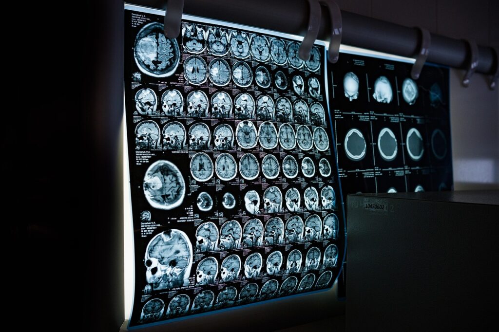
Computed tomography is one of the most heavily used diagnostic tools in modern medicine — roughly 90 million or more CT scans are performed in the United States each year (estimates vary and continue to rise). A CT scan uses rotating X-ray beams and computer reconstruction to produce detailed cross-sectional images of the body. It is fast, widely available, and especially valuable in trauma, stroke evaluation, and abdominal pain workups where speed matters. The trade-off is ionizing radiation exposure, which has prompted ongoing efforts to reduce dose without sacrificing diagnostic quality.
This guide explains what a CT scan is, how it works, how to prepare, and what the safety considerations are. It is general educational information, not medical advice. Whether you actually need a CT — and whether contrast dye is used — is a decision for your doctor based on your symptoms, your history, and what other tests have already shown.
How a CT Scan Works
A CT scanner consists of an X-ray tube and detector array that rotate around the patient as the table moves through the gantry. Hundreds of X-ray projections are acquired and a computer reconstructs them into thin cross-sectional images, typically 0.5 to 5 mm thick. Modern scanners produce images in seconds.
According to the RadiologyInfo.org resource maintained by the Radiological Society of North America (RSNA) and the American College of Radiology (ACR), CT visualizes both bone and soft tissue with high spatial resolution — better detail than a conventional X-ray, especially for soft tissues and blood vessels. The X-ray attenuation differences between tissues are displayed in shades of gray, with bone bright, fat dark, and soft tissues in between. Software can generate 3D reconstructions, vascular maps, and virtual endoscopy from the same data set. Unlike an X-ray film, CT leaves no radiation in the body after the scan is complete.
Common Reasons for a CT Scan
The NIH MedlinePlus resource and RadiologyInfo describe typical indications. CT is the first-line imaging test for suspected stroke (to rule out hemorrhage before clot-busting drugs), acute abdominal pain (appendicitis, diverticulitis, kidney stones), pulmonary embolism (CT angiography), trauma (rapid head-to-pelvis imaging), and lung nodule evaluation. CT is also routinely used to detect and stage cancers of the lung, liver, kidney, ovary, and pancreas, for treatment planning, to guide biopsies and drain placement, and for follow-up. It can even measure bone density in some osteoporosis protocols.
For kidney stones, low-dose non-contrast CT has become the diagnostic gold standard, replacing intravenous pyelography. CT colonography offers a non-invasive alternative to optical colonoscopy in selected patients. Where soft tissue contrast or radiation avoidance matters more than speed, an MRI is often the better choice. CT is not a general “scan everything” screening test for people without symptoms; whole-body scans marketed directly to consumers can turn up incidental findings that lead to more tests, cost, and anxiety without clearly improving outcomes.
Preparation and What to Expect
Preparation depends on the body region scanned and whether contrast is used. For most non-contrast scans, little or no preparation is needed. For abdominal CT with oral contrast, you may be asked to drink a barium or iodine-based solution over 1 to 2 hours before the scan. For IV-contrast scans, many facilities ask you not to eat for a few hours beforehand and want a recent kidney function test on file. Tell your doctor beforehand about any chance of pregnancy, recent illnesses, medications, and allergies.
You will change into a gown and remove jewelry and metal objects. You lie on the scanner table, which slides into the donut-shaped opening — much shorter and more open than an MRI tube, which is easier for people who feel claustrophobic. The technologist may ask you to hold your breath for 5 to 20 seconds during image acquisition. Most appointments take 5 to 30 minutes depending on the body region; the actual scanning time is often under a minute.
If IV contrast is used, you will feel a warm flush as it is injected and may sense a metallic taste briefly. These sensations are normal and resolve quickly. The scan itself is painless.
CT Contrast: Allergy, Kidneys, and Metformin
Iodinated contrast highlights blood vessels, organs, and abnormal tissue. Allergic-type reactions are possible: most are mild (a small percentage of patients), and severe reactions are uncommon (on the order of a few hundredths of a percent). Patients with a prior contrast reaction are typically premedicated with steroids and antihistamines or scanned without contrast, so it is important to tell the imaging team about any earlier reaction.
Contrast-associated kidney injury is uncommon in patients with normal kidney function but is a consideration in chronic kidney disease (historically flagged around an eGFR below 30, with extra caution in the 30–45 range). The ACR Manual on Contrast Media outlines current guidance, which favors hydration and using the lowest effective dose. Anyone with kidney disease, a single kidney, or a transplant should tell the team beforehand. Patients taking metformin are usually asked to follow their facility’s protocol around the time of contrast — in some situations metformin is briefly paused and restarted once kidney function is confirmed stable — because of the small risk of a rare metabolic complication if kidney function drops. Follow your own facility’s instructions rather than any generic rule.
Radiation Dose and Safety
Radiation dose is the other key consideration. A typical chest CT delivers roughly 5 to 7 mSv and an abdominal CT roughly 7 to 10 mSv — on the order of a few years of natural background radiation, which averages about 3 mSv per year in the U.S. A head CT is lower, around 2 mSv. The lifetime cancer risk attributable to a single medically indicated CT is small but not zero, which is why radiologists and ordering providers follow the “as low as reasonably achievable” (ALARA) principle, tailor the dose to the patient, and weigh CT against alternatives like ultrasound or MRI when those can answer the question.
The key point from both RadiologyInfo and the U.S. FDA is one of balance: when a CT is clinically indicated, the expected benefit of getting the right diagnosis usually outweighs the small radiation risk. The goal is to avoid unnecessary scans and to use the lowest dose that still answers the clinical question — not to refuse a scan your doctor believes you need. Modern protocols and dose-reduction software have cut typical CT doses substantially over the past decade.
What Happens After the Scan
You can usually resume normal activities immediately. If IV contrast was given, drinking extra water over the next day helps the kidneys clear the agent, though this is not strictly required for people with normal kidney function.
A radiologist reads the scan, typically within hours for inpatient and emergency studies and within 1 to 2 days for outpatient studies. Reports are sent to your ordering provider, who discusses the findings with you. Many systems now release reports through patient portals on the same day. Critical findings (active bleeding, aortic dissection, large pulmonary embolism) are communicated directly to the ordering clinician.
When results require urgent follow-up: If a CT identifies a finding such as appendicitis, dissection, large pulmonary embolism, or acute hemorrhage, you should expect immediate communication and direct admission or transfer for treatment. If you experience worsening symptoms (severe shortness of breath, chest pain, or severe abdominal pain) before results are available, do not wait — seek emergency care or call 911.
CT Limitations and Comparison With Other Imaging
CT excels at speed, bone detail, and acute trauma evaluation, but it has limits. Soft tissue contrast is lower than MRI, which matters for brain tumor characterization, ligament and meniscal tears, and pelvic disease. CT uses ionizing radiation, which is a more meaningful consideration for children, pregnant patients, and anyone needing repeated imaging over time. MRI uses magnets and radio waves instead of X-rays, takes longer, and gives superior soft-tissue detail; ultrasound uses no radiation and is often preferred first for gallbladder disease and for lower abdominal or pelvic pain in young women.
For pulmonary embolism, CT pulmonary angiography is the standard. For stroke evaluation, non-contrast CT is the first test, often followed by CT angiography or MRI. Lung cancer screening with low-dose CT in eligible adults — currently adults roughly 50 to 80 with a significant smoking history — is recommended by the USPSTF (confirm the current eligibility criteria, which are periodically updated).
Cost Considerations
Costs vary widely and are worth comparing. Without insurance, CT scans typically cost roughly $300 to $2,500 in the U.S. depending on body region, contrast use, region, and facility. Free-standing outpatient imaging centers usually charge less than hospital-based departments — sometimes far less for the same scan. With commercial insurance, out-of-pocket costs commonly run $100 to $700 after the deductible, and some plans require prior authorization for non-emergency CT. Emergency CT is bundled into the ED facility charge and can add substantially to a single visit. Because prices swing so much, it is reasonable to ask for the cash-pay or “self-pay” rate and to get the total in writing before you schedule.
Our healthcare costs guide covers how to shop for imaging, request cash-pay rates, and compare a hospital outpatient department with an independent center. The medical conditions resource library covers diagnostic workups across organ systems.
Frequently Asked Questions
How much radiation does a CT scan deliver?
Doses vary by body region and scanner protocol. A head CT is about 2 mSv, chest CT roughly 5 to 7 mSv, and abdominal CT roughly 7 to 10 mSv. For comparison, average annual background radiation in the U.S. is about 3 mSv. Modern protocols and dose-reduction software have reduced typical CT doses significantly over the past decade. Exact figures depend on your scanner and protocol — verify with your imaging center.
Are CT scans safe during pregnancy?
Doctors generally avoid CT in pregnancy when a good alternative exists, particularly for the abdomen and pelvis. Head and chest CT deliver very low doses to the fetus and may be acceptable when clinically necessary. Iodinated contrast crosses the placenta but is generally considered low-risk. If you are or might be pregnant, tell your provider so the plan can be tailored to you.
Can I eat before a CT scan?
For non-contrast scans, you can usually eat normally. For scans with IV contrast, many facilities ask you not to eat for a few hours, mainly to reduce nausea risk. For abdominal or pelvic CT with oral contrast, you may need to drink a contrast solution over 1 to 2 hours before the scan. Follow the specific instructions your imaging center gives you.
How is CT different from MRI?
CT uses X-rays and is fast, making it ideal for trauma, stroke, and abdominal emergencies. MRI uses magnets and radio waves, takes longer, uses no radiation, and provides superior soft-tissue detail. The right test depends on what is being investigated and how quickly answers are needed — a judgment your doctor makes with you.
Do I get to decide whether I have a CT?
You are always part of the decision, but whether a CT is the right test — and whether it needs contrast — is a clinical judgment your doctor makes based on your symptoms and other results. Good questions to ask include why CT was chosen over MRI or ultrasound, whether contrast is necessary, and whether an outpatient center could lower the cost.
The short version: A CT scan uses X-rays to make fast, detailed cross-sectional images and is the workhorse of trauma, stroke, and acute abdominal imaging. When a scan is clearly indicated, the benefit usually outweighs the small radiation risk, and radiologists follow ALARA to keep the dose down. Tell the team about prior contrast reactions, kidney problems, metformin use, and any chance of pregnancy. Prices vary a lot, so compare an outpatient center with a hospital and ask for the total in writing. Whether you need a CT at all is a decision for your doctor.
This article is general information, not medical advice. Talk with your doctor or radiologist about whether a CT is right for you, and seek emergency care for sudden severe symptoms.
The Bottom Line
A CT scan is fast, accurate, and widely available — qualities that make it the workhorse of emergency and acute care imaging. Useful conversations with your provider include why CT was chosen over MRI or ultrasound, whether contrast is necessary, what the radiation dose is for your scan, and whether outpatient imaging is an option to lower cost. For repeated imaging in younger patients, asking about cumulative radiation exposure and whether MRI or ultrasound could substitute is part of careful long-term care. When in doubt about your symptoms or your results, see a doctor.
Sources
- RadiologyInfo.org (RSNA and ACR) — Body CT (CAT Scan) and Contrast Materials
- American College of Radiology (ACR) — Manual on Contrast Media
- NIH MedlinePlus — CT Scans
- U.S. FDA — Computed Tomography (CT) and Medical X-ray Imaging radiation safety
- USPSTF — Lung Cancer Screening recommendation
Dose figures, screening criteria, contrast guidance, and prices can change; verify current recommendations with your imaging center and physician.